

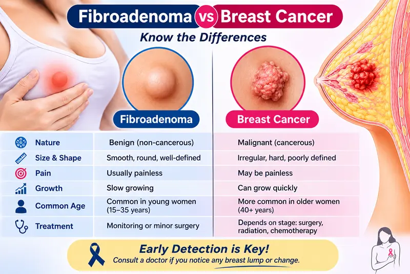
In the fibroadenoma vs breast cancer question, a fibroadenoma is a benign (non-cancerous) lump that is usually smooth, round and easy to move, while a cancerous lump is more often hard, irregular and fixed in place. But touch alone cannot separate them. Doctors confirm the diagnosis with a breast examination, imaging (usually ultrasound, with a mammogram depending on your age) and a needle biopsy. Any new or changing breast lump should be checked.
Finding a lump in your breast is frightening, and it is natural to fear the worst. Most breast lumps turn out to be benign, and fibroadenoma is one of the most common causes in younger women. This guide explains how the two differ, how doctors tell them apart, and when to get help.
Fibroadenoma vs Breast Cancer at a Glance
| Feature | Typical fibroadenoma | Features that raise concern for cancer |
| Nature | Benign (non-cancerous) | Malignant (cancerous) |
| Feel | Firm or rubbery, smooth, round or oval | Hard, irregular edges |
| Movement | Moves easily under the skin | May feel fixed to the skin or deeper tissue |
| Pain | Usually painless; sometimes mild tenderness | Often painless too, so pain is not a reliable guide |
| Common age | Most often 15 to 35, but any age | More common with increasing age, but can occur in younger women |
| Other signs | Usually none | Skin dimpling or redness, nipple turning inwards, bloody nipple discharge, lump under the arm |
| Ultrasound | Smooth, oval, wider than tall | Irregular or spiky edges, taller than wide, shadowing behind the lump |
| Confirmed by | Needle biopsy | Needle biopsy |
What Is a Fibroadenoma?
A fibroadenoma is a solid, benign breast lump made of glandular and connective tissue. It is often found by chance in the shower or during a breast self-examination, or on a scan done for another reason. Many women have one, but some have several, in one or both breasts.
Fibroadenomas are influenced by hormones. They may stay the same size for years, grow during pregnancy, or shrink after menopause. Some become smaller or disappear on their own. If you want to know what treatment can involve, see our page on breast fibroadenoma treatment in Delhi.
What Does a Breast Cancer Lump Usually Feel Like?
Breast cancer often appears as a hard lump with uneven edges that does not move freely. It may or may not be painful. Other possible signs include:
- Dimpling, puckering or redness of the skin
- A nipple that turns inwards or changes shape
- Bloody or clear nipple discharge from one breast
- A lump or swelling in the armpit
- A change in the size or shape of one breast
These are warning signs, not proof. Some cancers feel soft or smooth, and some benign lumps feel firm. That is why nobody, including a doctor, should rely on feel alone.
Why You Cannot Tell by Touch Alone
A smooth, mobile lump is reassuring but does not rule out cancer, and a hard lump is not always cancer. Fibroadenomas can be firm, and some cancers can be mobile. Self-examination is useful for noticing a change, but the diagnosis needs tests.
How Doctors Tell the Difference: Examination, Imaging and Biopsy
Doctors use a combination often called triple assessment:
- Clinical breast examination. The doctor checks the lump, both breasts and the lymph glands under the arms.
- Imaging. In women under about 30, ultrasound is usually used first, because dense breast tissue makes mammograms harder to read. In older women, a mammogram is often added to ultrasound. Ultrasound can also tell a solid lump from a fluid-filled cyst.
- Needle biopsy. A radiologist usually takes a small sample through a hollow needle under ultrasound guidance, using local anaesthetic. A pathologist then examines it. This is the only way to be certain of the type of lump.
Not every lump needs the same plan. Your doctor will decide based on your age, the appearance of imaging and your risk factors, such as a family history of breast cancer. Our department performs image-guided FNAC and biopsy procedures.
Can a Fibroadenoma Turn Into Cancer?
The risk of cancer developing inside an ordinary fibroadenoma is very low, and it is rare in women under 40. A simple fibroadenoma does not raise your future risk of breast cancer. Two points need care:
- Complex fibroadenoma. If the lump contains cysts, calcium deposits or certain overgrowth of cells, it is called complex. Studies suggest this type may carry a slightly higher future breast cancer risk, roughly twice that of women without one. This is a small increase and is usually managed with follow-up.
- Mistaken identity. A lump called a fibroadenoma on a scan occasionally turns out to be something else on biopsy, such as a phyllodes tumour. That is a good reason not to skip testing.
Having a fibroadenoma also does not protect you from breast cancer elsewhere in the breast, so new lumps still need checking.
Phyllodes Tumour vs Fibroadenoma
A phyllodes tumour is a rare breast tumour that can look and feel like a fibroadenoma. It tends to grow faster and be larger. Most phyllodes tumours are benign, but some are borderline or malignant, and even benign ones can come back after removal. A fine-needle sample cannot reliably separate the two, so a core needle biopsy, and sometimes removal of the whole lump, is used when a phyllodes tumour is suspected. A fast-growing or large lump should be reviewed without delay.
Observe or Treat? Pros and Cons
Once a lump is confirmed as a fibroadenoma, you and your doctor choose between watching it and treating it.
| Approach | Pros | Cons |
| Observation with follow-up | No procedure; many small lumps stay stable or shrink | The lump remains; repeat visits and scans; some people stay anxious |
| Image-guided removal or ablation | Small needle puncture, usually local anaesthetic, often less scarring | Only for suitable small lumps; residual tissue or regrowth possible; not available for every lump |
| Surgical removal | Whole lump removed; useful for large or fast-growing lumps and unclear biopsy results | Scar, longer recovery, anaesthesia risks, and a new lump can still form |
There is no single right answer. Small, biopsy-confirmed fibroadenomas that are not growing are often monitored, while growth, discomfort, unclear results or strong anxiety are common reasons to treat.
When to See a Doctor About a Breast Lump
See a doctor promptly if you notice:
- Any new breast lump, even if it feels smooth and movable
- A known lump that grows, changes shape or becomes hard
- Skin dimpling, redness or thickening
- A nipple that turns inwards, or nipple discharge, especially bloody
- A lump or swelling under the arm
- A strong family history of breast or ovarian cancer
Get urgent advice if a lump grows quickly. Do not wait for the next routine check.
Next Step
If you have been told you have a fibroadenoma, bring your ultrasound, mammogram and biopsy reports to your consultation. Our team can explain your options for breast fibroadenoma treatment in Delhi, including monitoring when that is the best choice. You may also like our guide to treating a fibroadenoma without surgery.
Call +91 99584 74870 or WhatsApp +91 99584 74870. Department of Interventional Radiology, Sir Ganga Ram Hospital, Old Rajendra Nagar, Delhi – 110060.
Medical disclaimer: This article is for general information and does not replace medical advice, diagnosis or treatment. Always consult a qualified doctor about any breast lump.
FAQ Section
How can I tell if a breast lump is a fibroadenoma or cancer?
You cannot reliably tell by feel. A fibroadenoma is often smooth, rounded and movable, while cancer is more often hard and irregular, but there are exceptions. A breast examination, ultrasound or mammogram and a needle biopsy give the answer.
Can a fibroadenoma turn into breast cancer?
The chance is very low. Simple fibroadenomas do not raise future breast cancer risk, and complex ones may raise it slightly. Rarely, a lump thought to be a fibroadenoma proves to be another tumour, so imaging and biopsy matter.
Is a hard breast lump always cancer?
No. Some benign lumps, including fibroadenomas, feel firm. Equally, some cancers feel soft. Any new hard lump should be examined and tested promptly.
Do fibroadenomas hurt?
Most cause no pain. Some women feel mild tenderness, which may vary with periods. Pain is not a reliable way to tell a benign lump from a cancer, because cancers can be painless as well.
Can a fibroadenoma and breast cancer occur together?
Yes, it is possible to have both, in different parts of the breast. This is why a new or changing lump should be assessed even if you already know you have a fibroadenoma.
What is a complex fibroadenoma?
It is a fibroadenoma with extra features under the microscope, such as small cysts or calcium deposits. It may be linked with a slightly higher future breast cancer risk, and doctors usually advise follow-up.
Is a biopsy always needed for a fibroadenoma?
A biopsy is the only certain way to confirm a fibroadenoma and is commonly advised, especially when the lump is not typical on imaging, is growing, or you are older. Your doctor will decide whether it is needed in your case.
What is the difference between a fibroadenoma and a phyllodes tumour?
Both can feel like a smooth, movable lump. A phyllodes tumour is rare, tends to grow faster and be larger, and can be benign, borderline or malignant. A core needle biopsy or removal helps tell them apart.
At what age are fibroadenomas most common?
They are most common between about 15 and 35 years of age, but they can occur at other ages too. A new lump in an older woman needs careful assessment.
When should I worry about a breast lump?
See a doctor for any new lump, a lump that grows or hardens, skin dimpling or redness, nipple changes or discharge, or a swelling under the arm. Early assessment brings peace of mind or earlier treatment.
4. References
- Mayo Clinic Care Network, fibroadenoma overview: https://mymlc.com/health-information/diseases-and-conditions/f/fibroadenoma?section=Diagnosis (symptoms, ultrasound, core needle biopsy, observation vs surgery).
- Altru Health System fibroadenoma overview (Mayo Clinic content): https://WWW.ALTRU.ORG/health-library/conditions/fibroadenoma (age range 15 to 35, ultrasound first under 30, phyllodes biopsy, complex fibroadenoma risk).
- Breast360, American Society of Breast Surgeons patient information on fibroadenoma: https://breast360.org/?p=210 (ultrasound appearance, biopsy as only definitive test, simple vs complex, risk).
- Medscape/eMedicine, fibroadenoma imaging overview: https://www.emedicine.com/radio/topic109.htm (complex or multiple fibroadenoma relative risk about twice; elastography; phyllodes features).
- AMBOSS, fibroadenoma: https://www.amboss.com/us/knowledge/fibroadenoma (biopsy indications; core biopsy preferred over FNAC when phyllodes suspected). Subscription site; verify against a primary source.
- NIH/PMC review on fibroadenoma management: https://pmc.ncbi.nlm.nih.gov/articles/PMC4562655 (malignancy risk below 0.3% and rare before age 40).
- Warning signs of breast cancer (skin dimpling, nipple changes, discharge, axillary lump): add a primary source such as the American Cancer Society or National Cancer Institute before publishing; this section is standard guidance but I did not retrieve a source for it.